In a detailed analysis of the requests sent to the Skinive bot, we noticed that people are very often worried not about the condition of a particular mole (pigmented nevus), but the quantity of the moles on their body. Moreover, with further analysis of the situation, it becomes obvious that there is a fact of special oncological alertness due to the appearance of more and more nevi. Many people see high oncological risks in this. But Is it really so dangerous? Is there a need to urgently visit a dermatologist or an oncologist?
In fact, there are even more options. In certain cases, you can safely stay at home without the risk of missing a serious skin problem. Spoiler alert: all you need to do is download a mobile application and conduct independent monitoring of the skin condition.
It is important to understand: if there are a lot of nevi on the skin, it does not mean that it is time to look for melanoma. On the other hand, when there are few nevi, this also does not mean that the skin condition can be ignored.
Indeed, in those people who have a lot of nevi, the risk of melanoma is slightly higher. And the problem is not even that a malignant transformation of the nevus can occur. Most often, the danger lies in the appearance of a new skin formation, rather than in the change of an existing one.
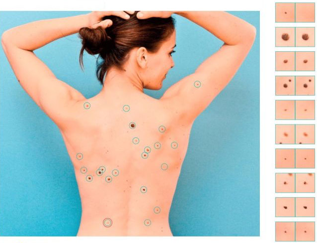
But both in the first and in the second case (with a large and small number of nevi) – the most optimal tracking option is to draw up a map of moles (this includes all other types of skin neoplasms), when each nevus, hemangioma, or any other skin pathology is considered in detail and is archived with reference to precise localization on the human body for the possibility of re-analysis and comparison in the future.
Why Mapping Moles?
The key goal of mapping moles is dynamic observation, which allows to timely identify the appearance of new formations on the skin or identify dangerous changes in them.
For people with few moles, mapping helps to notice changes in already existing neoplasms in time. The appearance of new mole in such people, as a rule, is always noticeable without the use of special devices (for example, there was one mole on the forearm, but now there are two: it is easy to notice the difference). Although there can be problems here, when the nevus appears in a visually inaccessible place (back, buttocks, back of the thighs).
As for people with multiple nevi (usually more than 50), mapping allows solving two main tasks.
The first task helps to keep track of the appearance of new pigmented neoplasms since it is quite problematic to do it on your own (for example, there are 7-8 nevi on the forearm, the appearance of another one may not be noticed).
The second task allows recording any visual changes in long-standing nevi. And since there are many nevi, here one cannot do without the help of photographic fixation and mapping. Moreover, in people with multiple nevi, as a rule, external changes in these neoplasms occur more often.
When and how often is skin mapping required?
Ideally, skin mapping should be done annually. However, if there are triggers, more often. They may include:
Active beach season (or any other long stay in the sun, especially with skin burns);
Presence of malignant tumors (especially skin, especially melanoma) in the history;
Pregnancy;
Taking hormonal or immune-inhibiting drugs (immunosuppressants);
People whose skin is in long-term contact with potential carcinogens due to professional activity.
And in the case when a person himself suspects the presence of possible dangerous changes in the skin. The frequency and regularity of mapping in such situations is determined individually at an appointment with a dermatologist or oncologist.
If a person is not in any risk group, it is enough to do mapping once a year in the spring season, before summer, so that later there will be something to compare with (if suddenly after summer there will be changes among skin neoplasms).
In autumn, immediately after the beach season, the mapping may not be informative due to the fact that there may not be any obvious visual changes provoked by the summer sun by this time.
How to do a mole map?
In fact, the process of creating a mole map is quite simple. Under this term lies a banal photographing of individual areas of the skin, which together should make up the entire body as a whole. After that, each neoplasm is examined separately for the presence or absence of risks, the exact location, changes in comparison with previous images (if there are archived images) are recorded.
Given the uncomplicated technology, this procedure can be carried out at home using improvised means. Fortunately, modern mobile phones are equipped with quite good (and sometimes very good) cameras. Well, or you can use a professional technique. Processing photos on a PC with an indication of the neoplasms localization should also not be difficult. In principle, this approach can solve some problems, for example, to fix the appearance of a neoplasm where it did not exist before. Or, note changes in an already existing neoplasm, but these changes should be sufficiently pronounced (which does not always correspond to the timeliness of seeking medical help).
It is also necessary to understand the presence of a number of other important points:
The process of “home” mapping is not automated, everything is done manually, which requires a lot of time and effort;
The obtained images are not standardized: different distances from the camera to the skin, different lighting, there may be different cameras, different image quality during repeated mapping;
The inability to perform a dermatoscopic image (you can only get a macro photo, a photo of a neoplasm under a magnifying glass is not a dermatoscopy);
Lack of medical opinion.
Taking all this into account, skin mapping should only be trusted by professionals and professional techniques.
Tools for skin lesions mapping.
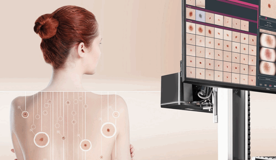
Usually, mapping of skin neoplasms is performed in health care facilities by specially trained doctors using special equipment. In the office, there is a stand with one or several cameras that take pictures of a person according to a predetermined algorithm. Such a device is also equipped with an electronic dermatoscope, with the help of which skin neoplasms are examined in detail under high magnification. There is also artificial intelligence that helps doctors to make the right decision. All this information is recorded in the patient’s personal account and stored until the next visit. Today, this technology is considered the benchmark for skin mapping.
Of course, the reliability and result of such a procedure should not raise any doubts. But here comes another problem: accessibility. The cost of such installations is estimated at tens of thousands of dollars, which not every doctor and not every medical center can afford, where dermatological and oncodermatological patients are consulted. Accordingly, not even in every large city, you can find such a service. And everyone needs to watch out for skin neoplasms.
An alternative way to perform skin mapping can be dedicated mobile applications for smartphones, such as Skinive.
Download Skinive MD app for iPhone and Android
The main advantages of this approach are accessibility and mobility.
The procedure can be carried out in any office (as opposed to stationary installations, which are tied to a specific office), where there is sufficient lighting and the patient feels comfortable. The mapping process itself is controlled by artificial intelligence: each neoplasm that the neural network was able to identify is saved and linked to a virtual 3D model of a person. After the end of the skin scan (also carried out according to a special standardized algorithm, which must be familiarized with before starting the scan), all neoplasms that the neural network previously recognized as potentially dangerous are highlighted for the doctor. After that, each such neoplasm undergoes a more detailed check with a neural network, on the basis of which the doctor makes his conclusion. All results are stored in the patient’s personal account and are available for comparison at the next mapping.
It is difficult to disagree with the fact that this approach to compiling a mole map most fully meets modern requirements:
It is not tied to a specific doctor’s office;
Available to a wide range of doctors and institutions;
The results can always be at hand or, if necessary, available to other doctors (joint consultations, patient transfer, training).
Moreover, in order to carry out mapping in this way, special in-depth knowledge in dermatology or dermato-oncology is not always required. This technique can be considered as a screening test by general practitioners or other non-skin specialists to properly route the patient.
Thus, our health and the health of our skin, in particular, are truly in our hands.
Artificial intelligence has been used in health care for many years: to analyze medical images (X-rays, MRI, CT, etc.), to create expert systems and virtual medical assistants.
But however impressive it may be, AI cannot replace human experience. Ultimately, it is the doctor who determines the next steps. Artificial intelligence is a virtual assistant which can improve diagnostic accuracy and save the doctor’s valuable time.
Dermatologists have a tough job. In order to correctly determine the cause of skin disease and successfully prescribe the treatment, dermatologists monitor the entire patient’s body, evaluate its condition and, if necessary, use additional diagnostic techniques, such as biopsy.
Skinive for medical professionals.
At Skinive we see the great potential of our solution use not only by dermatologists but also by all specialists who directly or indirectly deal with skin health issues, such as cosmetologists, nurses, general practitioners, allergists, venereologists.
1. How to take off the pressure the dermatologists?
For general practitioners (therapists), it is even more difficult to determine the nature of neoplasm – as this is not their specialty. According to the statistics, about 20 percent of all the initial visits to therapists refer to skin health issues and almost all of these patients are sent to dermatologists: those who really need treatment, and those who don’t have any problems. This creates a very high workload for dermatologists. Healthy patients also take the time of a dermatologist and prevent them from concentrating on patients who need help.
2. The use of Skinive by primary healthcare specialists.
Skinive screening helps those who are not well trained on skin conditions or do not have deep knowledge in dermatology to assess the initial risks.
The Skinive Express Tests (fast-checks) allow doctors to take images of suspicious neoplasms and get a preliminary conclusion from the Skinive algorithm in 30 seconds. After you send a photo to the Skinive application, you will get a preliminary result based on the differential diagnostic method (excluding any medical condition that is not appropriate for any of the facts or symptoms that the patient may have, which should ultimately consolidate the diagnosis to a uniquely probable disease).
The differential diagnosis of the Skinive algorithm includes:
3 hypothetical diagnoses detected by our algorithm between the analyzed image and the database;
Percentage of similarities detected by the algorithm for each of the variants of the assumed similarities (The higher the percentage, the better the similarities);
Risk assessment: Low, Medium, High. Benign neoplasms – Low risk. Acne and HPV – Medium risk, neoplasms with high malignancy risk or malignant tumors – Hight risk.
Recommendations for a specialist routing (cosmetologist, dermatologist, oncologist, etc.)
Important!
In order to avoid diagnostic errors, we recommend routing patients to a specialist in all cases where at least one of the estimated three variants indicates on a medium- or high-risk level and more than 25% of similarity detected.
3. How do we help dermatologists to examine skin?
One of the biggest problems in dermatology is the difficulty of visual diagnosis due to the absence of a large clinical case database. This is especially relevant to young professionals with a lack of medical experience.
Experienced dermatologists are exceptionally good at finding potentially dangerous skin tumors that stand out in the background of others, but it can be difficult to track changes (if there are any). Skinive provides dermatologists with a tool for the rapid tumors mapping and the use of this data for observation over time.
Skinive AI will help you to improve your medical practice with a wide array of benefits:
Patients management: quick patients profile creation and user-friendly search engine;
3D Skin map allows to speed up the process of documentation: it is enough to mark the location of the photographed skin area on a 3D human body model.
Skinive AI Smart Camera makes high-quality photos with smartphones, allowing dermatologists to see and evaluate skin problems.
Secure data storage to track changes over time and an easy and convenient way of taking notes.
A second opinion from AI: differential diagnosis of Skinive AI allows to exclude the unsuitable facts or disease symptoms, thus, simplifying the diagnosis.
Dermatological Atlas provides quick access to detailed disease descriptions, diagnostic methods and treatment options.
4. Carrying out Skinive photo examinations.
This sketch illustrates the Skinive photo examination process.
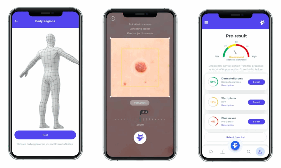
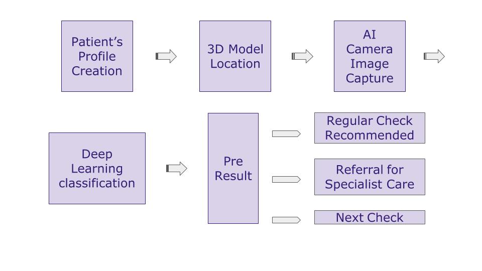
Create a new Patient Profile or use Search to find an existing one;
Mark the tested body area on the 3D body map;
Take a snapshot of the affected skin with the Skinive app. Keep the smartphone camera at the right angles. Center the object. Maintain a minimum focal distance (10-15 cm) from the object. Use zoom if necessary;
Get a preliminary report from the top 3 results: the higher the Top – the higher the probability of this particular pathology. For dermatologists and oncologists, there is a possibility to choose the most reliable result from three proposed variants, or to choose another nosology from the additional list if it does not appear in the proposed Top 3 list;
After choosing the desired nosology, the result will be saved in the application.
The result of the photo examination includes:
patient data and time of photographic examination;
original photo with a neoplasm, the segmented area image with a lesion and a visualization of the dysplasia level;
Class and type of nosology: presumed diagnosis and reference to an article on a given nosology in a dermatological atlas;
Risk Level (High, Medium, Low);
Your comments.
You can change the probabilistic diagnosis after having the results of the additional diagnosis (biopsy). If the risk of the disease is low but the observation over time is required – schedule the next photo-examination.
How does Skinive analyze images?
Skinive uses a deep machine learning algorithm (AI-algorithm). The human ability to learn from examples and experiences has been transferred to a computer. For this purpose, the neural network has been trained using a dermoscopic imaging database containing tens of thousands of examples that have confirmed diagnosis and assessment by dermatologists.
The AI is able to distinguish between benign and malignant tumors, similar to the ABCDE rule (5 main signs of oncology: asymmetry, boundary, color, diameter, and change over time). The difference between them is that the algorithm can analyze thousands of features, but not only 5 of them. Of course, only a machine can detect that amount of evidence.
Due to the productive cooperation with doctors, the quality of the Skinive algorithm performance is constantly being improved. Based on growing experience and its own autonomous rules, the AI is able to distinguish between benign and malignant tumors, find risks of human papillomavirus, and classify different types of acne…
Use Skinive in your medical practice and join Skinive network!
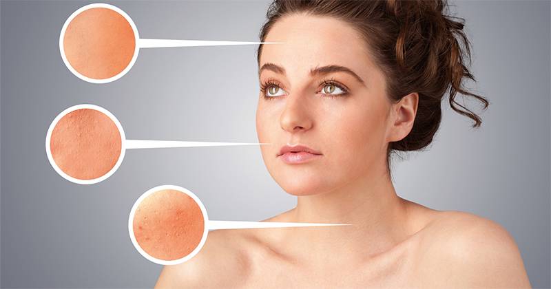
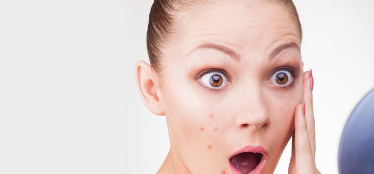
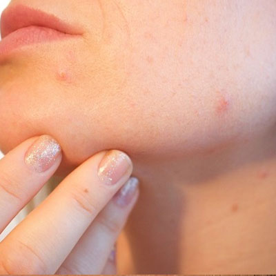
Acne is a broad term that encompasses a variety of skin ailments. It’s a condition that involves much more than an occasional pimple or zit. Acne comes in varying severity and it affects everyone differently. In order to successfully treat your acne, it’s essential to recognize and diagnose the different acne types you are struggling with (you may have more than one). Bear with us, these aren’t the nicest names or descriptions.
Types of Acne – What Type of Acne Do I have?
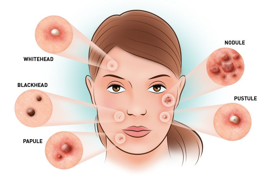
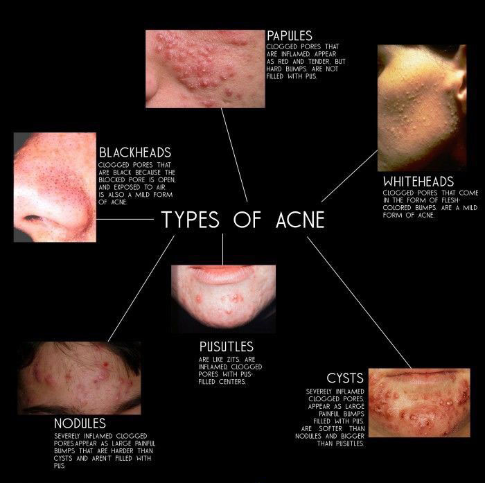
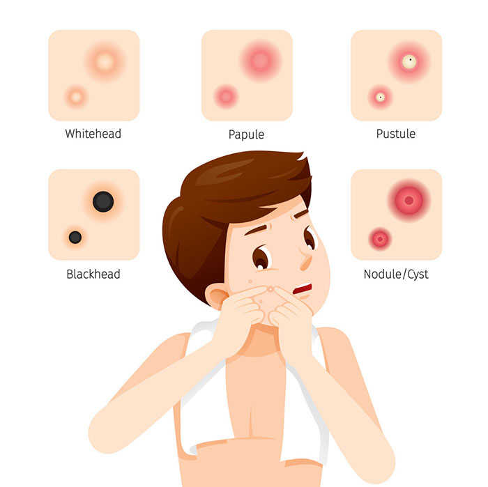
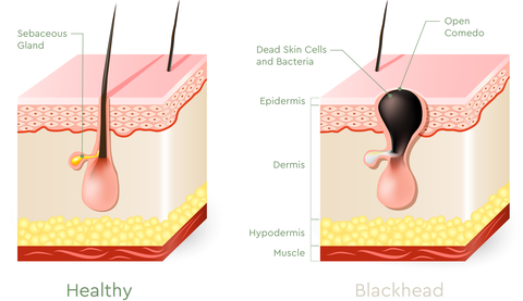
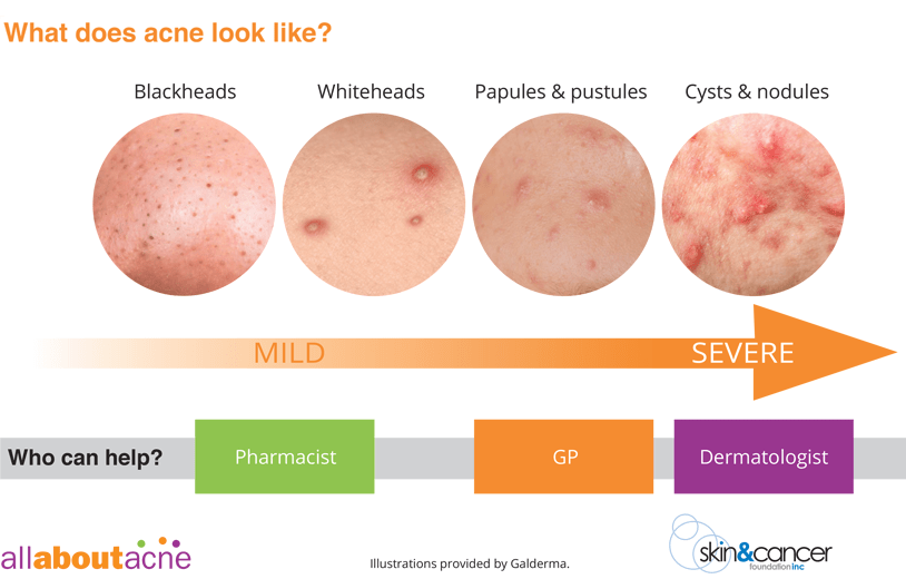
Acne Vulgaris presents different types of acne lesions: blackheads, whiteheads, papules, pustules, nodules, and cysts:
Blackheads: These are uninfected, clogged follicles that appear as a dark bump on the skin.
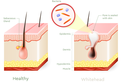
Whiteheads: Whiteheads are clogged follicles covered by a thin layer of skin that appear as white bumps or spots.
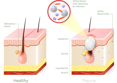
Papules: Papules are inflamed lesions that may appear red, and can be sensitive and painful.
Pustules: Pustules are inflamed lesions that are generally pus-filled. They may appear white or yellow, and popping pustules can lead to acne scarring.
Nodules: Nodules are a severe form of acne lesion that develop under the skin; they don’t generally contain pus but are hard to the touch.
Cysts: Cysts are a severe form of acne lesion that are inflamed and filled with pus. They are generally painful and require professional medical treatment.
Beyond Acne Vulgaris, there are different kinds of acne conditions, including Acne Fulminans and Acne Mechanica.
Types of Acne Images
Acne Fulminans: This is a rare but very severe form of acne that comes on abruptly in adolescent male, characterized by inflammatory nodular acne on both the chest and back. It can result in severe scarring and painful joints, along with other health issues.
Acne Mechanica: This form of acne is triggered by excess pressure, heat, and friction on the skin, and is often found among athletes and those already prone to acne breakouts. It’s characterized by small bumps that can range from tiny comedones to inflamed lesions.
How Acne Develops
Acne is the emergence of infected or inflamed sebaceous glands in the skin. Acne Vulgaris—the medical name for common acne—forms when hair follicles become clogged with excess sebum, bacteria, and dead skin cells. This results in a comedo, also known as a clogged pore. Acne Vulgaris can be commonly found on the face, back, and chest, as these areas of the body tends to be saturated with pilosebaceous units, but may be found on other areas such as your booty. As skin cells regenerate, old cells slough off and. If these dead skin cells aren’t flushed from the pores and manage to combine with sweat and oil, they’ll be trapped and result in the formation of a pimple.
Our pores are connected to a system of oil glands directly beneath the skin. Within these oil glands, sebum is produced. This substance is meant to keep our skin moisturized and healthy, but excessive production of this oil can lead to acne; as oil is pushed through the follicle, should it attach to dead skin cells or a bacteria, it can clog the pore and create a plug in the skin. Sebum will continue to build up behind the plug, resulting in a comedo that may turn into one pimple.
General causes
There’s a plethora of myths about the causes of different types of acne, many of which are untrue and baseless. You might have been told to wash your face more, or cut down on your chocolate-eating habits, perhaps even told to get some more sun for your skin condition. None of these common acne myths are true. Poor hygiene doesn’t cause acne, chocolate won’t make you break out, and sun exposure can actually damage your skin and make breakouts worse. Acne is caused mainly by genetics and hormones, and there are many factors that can exacerbate this skin condition.
Hormonal Changes
Acne Vulgaris generally develops during the teen years, when the onset of puberty causes the hormonal level to fluctuate. As hormone levels rise, especially testosterone, the skin glands begin producing larger amounts of sebum. What causes oily skin? Sebum, which can lead directly to acne bumps or breakouts.
Puberty isn’t the only time we experience hormonal changes. Women experience regular variations in hormone levels, specifically that of estrogen and androgen. Men also experience hormone level fluctuations, especially in their teens, but this usually mellows out by adulthood.
Hereditary Factors
Acne is also a result of hereditary factors. Children whose parents have dealt with acne are more likely to struggle with this skin condition. While it’s not a genetic disease, hereditary components have been linked to the presence of this skin condition. Genetics have a huge bearing on how your immune system works; say two individuals experience the same bacterial infection. One might react with painful, pus-filled nodules, while another’s skin may only result in the formation of blackheads. Similarly, one person may have more sensitive skin that grows raw and inflamed more often due to their genetic predispositions. Ever asked yourself: Why do I have dry skin? Family history can have a lot to do with the way your skin looks and feels, and is usually a good indication of whether or not an individual will deal with acne breakouts.
Stress
Stress Acne
While stress doesn’t directly cause acne, it can trigger or exacerbate a breakout. We see this occur in students across the globe when they’re handling stress during finals. When you’re stressed, your body releases cortisol and androgens. When these hormones fluctuate, your skin secretes more oil, which can bring on a breakout or worsen pre-existing pimples. If you find that your skin is particularly sensitive, be sure to integrate healthy skin care tips to counteract the inevitable stress in your life.
How Acne Types are Categorized
Non-Inflammatory Acne
Acne is typically categorized into two main types: non-inflammatory and inflammatory acne. Although there are two primary categories, there are many different types of acne which we’ll discover later on.
Non-inflammatory acne is characterized by comedones, which can be open or closed. These comedones are more commonly known as whiteheads and blackhead and are generally referred to as different types of pimples.
Blackheads
If the pore stays open on the surface of your skin, it’s considered an open comedo, also known as a blackhead. Because the pore is open, the sebum inside the pore oxidizes upon contact with the air; this hardens the sebum, and once the air mixes with melanin skin pigment, the blackhead forms a dark coloration that can appear black, brown, or gray. Blackheads can usually be found on the nose, chin, and forehead, but might also be found on the chest, back, and arms. Blackheads tend to be rampant at the onset of puberty and during times of hormonal change.
Whiteheads
If a pore is not open and covered by a thin layer of skin, sebum and dead skin cells accumulate, resulting in a thick substance that remains stuck under the skin, forming a plug. While these are not usually infected, bacteria can affect the skin cells surrounding the pore. Whiteheads don’t usually last as long as blackheads, with an average cycle of a week. Whiteheads tend to be found on the face, but may also be observed on other parts of the body.
Self-extraction of comedones can cause more harm than good. The follicle walls in blackheads and whiteheads can be ruptured quite easily, and this rupture allows bacteria to enter the cells and leads to inflamed acne. That’s why it’s essential to avoid picking the skin, as this can lead to rupturing.
Inflammatory Acne
Inflammatory breakouts are a result of P. Acnes bacteria infecting the pore and causing an infection, and can often be more difficult to treat. There are four types of inflammatory acne, sometimes commonly referred to as different pimple types: papules, pustules, nodules, and cysts.Papules
Papules are small, raised, solid pimples that don’t contain pus and are usually the first type of inflammatory acne to affect the skin. Papules don’t display a visible pore and tend to be red in color and surrounded by swollen and inflamed tissue. They may be tender to the touch.
Pustules
Pustules are pimples that are noticeably white or yellow in the middle and are filled with pus. The marked difference between papules and pustules is that pustules contain white blood cells. When your immune system attempts to fight bacteria that’s made its way into a ruptured follicle, it will set off a buildup of white blood cells, resulting in pus production. The skin around pustules tends to be tender and inflamed.
Nodules
Acne nodules appear during the latter stages of a breakout and are usually seen in severe cases of acne. They present as large, tender bumps underneath the skin’s surface and can feel hard and stiff to the touch. Similarly to papules, nodules form from a buildup of bacteria, skin cells, and sebum in the follicles, but this formation is rooted deeper in the skin. This deep buildup can cause tissue damage to the skin and prompt the immune system to begin an inflammatory response.
While they look similar to papules, nodules are much bigger and more painful. They do not contain any pus, but can remain buried within the skin for a long time. They may be dormant and rear up in occasional bouts. If squeezed or ruptured, these nodules can spread over a larger area of the skin and cause deep infections. These lesions can cause damaging and permanent scars to the skin.
Cysts
Cystic acne is one of the hardest types of acne to successfully treat. Unlike pimples, acne cysts form deep below your skin tissue and do not come to a head. Cysts are similar to nodules, except for the presence of pus. Cysts are almost always painful to the touch and can cause great discomfort, and they don’t necessarily look like a pimple. They may just appear as a large, swollen, and red lump on the skin. They may occur independently, or you may find your skin has large clumps of cysts grouped together.
Cystic acne is less prevalent; where general acne affects 70 out of 1,000 people, cystic acne affects 2 in every 1,000 people according to Medical News Today. This severe form of Acne Vulgaris usually requires the help of a dermatologist in conjunction with self-care and preventative measures.
Inflammatory acne takes much longer to heal and can have permanent effects on the skin, so it’s important to determine the best treatment methods for your skin condition.
Sebaceous Filaments
If you’ve noticed a plethora of small grayish dots appearing across your face, especially on your nose or chin, you might incorrectly label them as blackheads. Those are actually not blackheads, they are what’s known as sebaceous filaments. These are naturally occurring hair-like formations that follow the flow of oil along the inside of a pore. If you have oily skin or larger pores, they’re more noticeable. These become visible as a pore is filled with sebum. While blackheads also begin as pores filled with sebum, it’s only once a pore is plugged with dead skin cells that it becomes an actual comedo.
It’s easy to see the difference between blackheads and sebaceous filaments when it comes to extraction. When extracted, a blackhead has a dark, hardened, plug-like appearance. Sebaceous filaments are expelled in a flowing stream of sebum that takes on a waxy consistency. You can also observe a difference in the colors of these lesions; a blackhead will be quite dark on the surface of your skin, where sebaceous filaments may be yellow-tinged, gray, or take on the hue of surrounding skin.
Even when these filaments are extracted, they generally refill with sebum within 30 days. There’s no way to completely rid the skin of these sebaceous filaments, but keeping your face clean with a regimen of regularly treating acne can help minimize their appearance and keep those pores cleaned out.
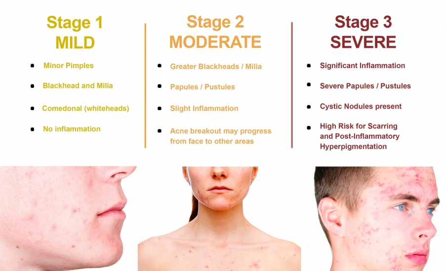
Severity Grades
When it comes to acne diagnoses, Acne Vulgaris is measured on grades of severity, and can be diagnosed in four different classifications.
Grade I
Patients diagnosed with Grade I acne generally exhibit mild forms of this skin condition. Their skin may have whiteheads, blackheads, and small pimples. There’s not usually inflammation present with this type of acne breakout, and mild cases like these can often be self-diagnosed. Usually, this type of breakout occurs on the nose and forehead and most often appears in adolescence. Adults may also experience this type of acne, usually with blackheads on the nose or forehead.
Grade II
Moderate acne falls under a Grade II severity qualification. Those with this grade of acne will likely exhibit a plethora of comedones on the skin. There will be more frequent breakouts, likely accompanied by pustules and papules that may be more sensitive to the touch.
Grade III
This grade of acne is accompanied by a large amount of inflammation in comparison to the lesser grades. The skin will exhibit numerous papules and pustules, and there may be nodules present.
Grade IV
This is the most severe form of acne, and will see skin filled with nodules, cysts, pustules, and papules. With this severity grade, acne tends to be spread across more than just the face; the back, chest, neck, and buttocks may also experience frequent breakouts. Acne of this severity can cause significant scarring and may disfigure the skin if left untreated. According to the American Academy of Dermatology, oral isotretinoin is the only medication approved to treat severe cystic acne.
What Does Acne look like
It’s important to seek out a dermatologist’s advice if you believe your acne is severe. The more severe the grade of your condition, the higher the risk of permanent skin damage, so it’s essential to secure the best treatment and take preventative measures to circumvent this.
Other Types of Acne
There are other different types of acne that are not considered Acne Vulgaris. They can look similar and exhibit comparable symptoms, but they tend to be much more difficult to treat and alleviate. If you are struggling with severe acne-like symptoms, it’s important to determine exactly what is causing your skin condition in order to access proper treatment.
Acne Conglobata
This is an uncommon and severe form of acne that’s characterized by painful abscesses and irregular scars, and features a large number of inflamed comedones, nodules, and draining sinus tracts all connected underneath the surface of the skin. It can begin with blackheads found in groups of two or three, and might be located on the face, chest, arms, buttocks, or neck. Pimples form around these blackheads and become engorged with pus. These will continue to grow until so filled with pus that they rupture. The pus within these nodules tends to be foul smelling. Once these have ruptured, the nodules may fuse together to create larger shapes and scab in the middle.
Generally, this skin condition affects men, as it has been associated with higher levels of testosterone, but women can also struggle with Acne Conglobata. The cause of this type of acne is unknown, but it tends to affect individuals between the ages of 18 and 30, and can persist until an individual reaches their 40s. Even after clearing up, this type of acne can cause permanent, significant damage to the skin.
Acne Fulminans
Acne Fulminans is a severe form of Acne Conglobata that is accompanied by horrible systemic symptoms. While very rare, this condition almost always affects adolescent males. This type of acne can come on suddenly and severely, with inflammatory and ulcerated nodular acne appearing on the chest or back. Also known as Acne Maligna, this condition can actually begin with pain and swelling in the joint and turn into nodular acne. These ulcerative acne lesions can bleed and crust over. In extreme cases, patients have experienced loss of appetite and weight loss, along with the potential for an enlarged liver or spleen.
Gram-Negative Folliculitis
This is actually a bacterial infection that closely mimics acne, but in reality is merely a pustular rash. Doctors can diagnose this condition with a Gram stain, but this condition can be difficult to treat, as the types of bacteria present in this condition don’t respond to most antibiotics used to treat severe acne. This infection can arise from continued use of certain antibiotics prescribed to treat acne; this is because the body builds up an immunity to these antibodies over time, resulting in worse acne flares. Gram-Negative Folliculitis is rare; it is estimated that four percent of individuals who have inflamed acne will develop this condition.
Pyoderma Faciale
Pyoderma Faciale can mimic both acne and rosacea, but it is neither. It was actually formerly classified as a variant of rosacea but has recently been reclassified as its own diagnostic entity. This painful skin condition only afflicts women, usually between the ages of 20 and 40 years old, and its onset is quick and immediate. Individuals with this skin condition will exhibit cysts, pustules, and nodules seemingly overnight. There are usually no comedones present with this condition on its own, but it is known to accompany cases of Acne Vulgaris. Usually, this skin condition lasts no longer than a year and is not associated with the production of oil in the skin. This is a rare condition and is usually best treated with medication.
Achne Mechanica
Acne Mechanica is a specific form of acne that is triggered by excess heat, friction, or pressure on the skin. It usually affects younger athletes, soldiers, and students. Anything that traps heat against the body, whether it’s clothing or athletic equipment, can elicit a breakout. Heavy gear or bulky clothing worn tightly to the skin can also affect the prevalence of this skin condition. Those prone to body acne are more likely to be affected by this condition. This condition can be prevented with absorbent material and showering immediately after activity.
Emotional Impact
No matter the severity or type, individuals dealing with any type of acne breakout and other skin conditions often struggle with emotional effects as a result. For teenagers early in their social development, skin ailments such as acne can cause self-esteem issues and in severe cases, depression. Even with the best young adult acne treatment put to use, acne breakouts and scarring can mar a young person’s self-image as physical appearance becomes important to feelings of self-worth.
The threat of emotional damage doesn’t stop in adolescence, however. Adults who deal with different kinds of acne can also face negative emotional impact as a direct result of their skin condition. One study performed by the Indian Journal of Dermatology found that adult females with mild to moderate acne experienced higher levels of emotional and social impairment.
Whether you deal with the occasion blackhead or have been experiencing severe and painful breakouts, acne is a daily struggle for many in the United States, with millions across the country struggling with this painful skin condition. When acne flares, causing physical discomfort and pain and negative psychosocial effects, treatment is vital. Be sure you know how to figure out your skin type as this understanding can aid you in any treatment you pursue. Narrowing in on the acne types you struggle with will enable you to select an effective treatment that clears your skin and prevents future breakouts.
Key Takeaways
There are two main types of acne: non-inflammatory and inflammatory.
Acne can cause significant negative psychological effects.
Sebaceous filaments are often mistaken for the presence of blackheads.
Dermatologists diagnose Acne Vulgaris in four grades of severity, ranging from mild to severe.
There are more severe skin conditions that are commonly mistaken for common Acne Vulgaris, including Acne Fulminans, Pyoderma Faciale, and more.
Left untreated, acne can leave permanent scars, so it’s essential to begin treatment as soon as possible.
“Discovering a skin cancer early can make the difference… between life and death.”
The difference between life and death…” Wow. As frightening as that might be, never underestimate the power that comes with knowing the techniques for early detection. Remember, skin cancer is almost always curable — if it’s detected and removed early.
Knowing the facts enables early detection
After reviewing your risk factors in Skin Cancer Part 1,1 and how to reduce those risks in Skin Cancer Part 2,2 you are now ready to learn how to perform regular and thorough skin checks — on yourself, your spouse, and your children. Remember: You are your very best resource for detecting skin cancer in its early stages.
Exams, both personal and professional, are life-and-death important
Besides skin checks at home, schedule regular and as-needed medical skin exams for yourself and your family — especially if any of you is at increased risk for skin cancer.
Annual skin exams by a health care professional have been shown to reduce deaths from melanoma by half.3 If your skin exam wasn’t performed by an actual dermatologist, follow up with one for an evaluation of any suspicious findings. Expect your dermatologist, an MD with specialized education and experience diagnosing and treating skin cancers, to listen carefully to your concerns, perform a thorough skin examination, and clearly communicate findings and recommendations for follow-up.
Monthly personal and annual professional skin checks improve your own, your spouse’s, and your children’s chances for early detection, treatment, and survival of a skin cancer diagnosis.
Do I really need to check my children for skin cancer?
Yes, absolutely
The following facts provided by Dana-Farber on childhood skin cancer are clear: Although rare, children dodevelop skin cancer — and it’s almost always melanoma — the deadliest of the three most common skin cancers.4
The risk of melanoma increases for those children with the same risk factors as adults (fair skin, sunburns, moles, family history). Moreover, the biggest increase in melanoma diagnoses is in girls 15–19 years old, most likely related to their sunbathing and tanning bed habits as well a seeming reluctance to wear protective clothing.
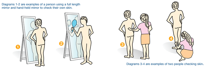
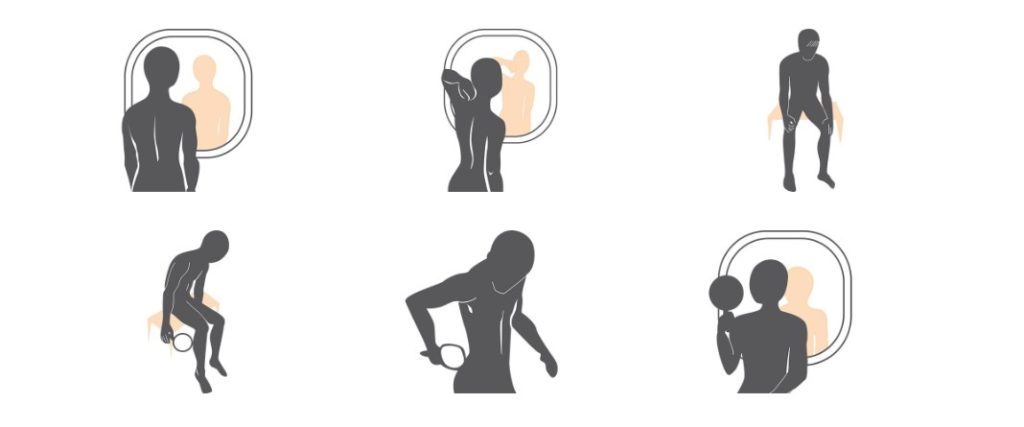
Follow the step by step instructions in this gallery to examine your skin. Take Smartphone shots to use Skinive
If your child falls into an increased risk category, perform their monthly skin checks and schedule them for annual and as-needed skin examinations by a pediatric dermatologist.
Don’t be frightened — you can do this. Read on for the additional information you’ll need to practice good stewardship of your own and your family’s skin health:
what a suspicious mole or lesion looks like; and
how to perform a skin check.
When is a mole or lesion suspicious?
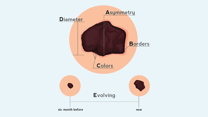
ABCDE Skin Self Examination
One or more of the following characteristics is highly suggestive of a melanoma, the most lethal skin cancer, and should be immediately evaluated by a dermatologist or, if a child, a pediatric dermatologist. To help you remember these, think ABCDE.
A=Asymmetry One half is a different shape from the other.
B=Border The edges are irregular.
C=Color There are variations in color (tan, brown, black, red, white) within the borders. Children’s melanoma is more likely to be whitish, yellowish, or red and somewhat wart-like in appearance 5
D=Diameter This characteristic is less definitive — while often larger than a pencil eraser, they can be smaller.
E=Evolving The size, shape, color is changing or there is itching, bleeding or difference from your other moles or lesions.
Of all these characteristics, an evolving or changing mole or lesion is the most suggestive of melanoma. And you are the best person to spot these changes early. Check your skin regularly!
The characteristics of basal cell and squamous cell lesions (the most common and second most common skin cancers) may be only a growth, bump, or sore that is unusual in appearance or does not heal within a month.
The slideshow Precancerous Skin Lesions and Skin Cancer can help you recognize what you might find during your skin checks.6 For anything that makes you suspicious of skin cancer or pre-cancer, make an appointment with your dermatologist for an immediate evaluation.
How to perform a skin check
What you’ll need
good lighting;
a comb or hair dryer to part hair;
a mirror if you’re checking your own scalp and back surfaces;
a small flexible plastic or paper millimeter (mm) ruler;
a flashlight; and
a means to document what you find (paper, computer, or smartphone app).
Photographing and dating lesions and regions (your back, your chest, your thigh, etc.) is very helpful in documenting your skin status.
Measure lesions and moles using a millimeter (mm) ruler
from side-to-side first;
then from top-to-bottom; and finally
the distance, if any, each is raised on the skin.
What to check
Whether performing skin checks on yourself or family members, leave no areas unexamined. This includes: the scalp and face; the fronts, tops, and backs of ears; around the neck; tops of shoulders; full front and back; as well as both sides from armpits to ankles.
Also check: the sex organs; fronts and backs of arms and legs; tops of hands and feet; and between all fingers and toes.78 The palms and soles as well as under and around all nails should be checked by everyone — but are especially important to dark complexioned persons as they are more likely to develop a virulent strain of melanoma in these areas.9 Did I forget anything? Check that too.
The baseline and follow-ups
The first full-body skin check can take quite a while as you are creating a baseline. Try dividing it up: all fronts, hands, and feet one day; then the scalp, all sides, and backs on another (perhaps when you have help).
Follow-up checks — when you’re looking for changes to the already documented moles and lesions or the emergence of new ones — should take only 10–15 minutes.
Keep track of what you find
As you’re checking, record each finding for each family member. You can use preprinted forms or create your own. And, not surprisingly, there’s an app for that.
As your children mature, have them join you, first checking and later recording easy skin findings. This will teach them the habit, the skills, and the importance of regular skin checks.
The skcin.org website provides a useful form to use as you inspect and document lesions and moles.10
Minimize the impact of skin cancer on yourself and your family
The importance of protecting yourself and your family from skin cancer — as well as assuring its early detection and successful treatment — cannot be overstated.
As part of your protection plan, you should:
mandate, demonstrate, and facilitate avoidance of ultraviolet ray exposure;
perform thorough monthly skin checks;
schedule annual medical skin examinations; and
schedule immediate dermatologic evaluation of any suspicious moles or lesions.
Early discovery of skin cancer can be the difference between office-based surgery and far more invasive treatment, even between life and death. Choose life.
Good stewardship
As believers, we know that God is the creator and possessor of all things in Heaven and on Earth (Genesis 1:1–27 NKJV). And that we are to be good stewards of His possessions, including His most precious possession, our bodies. “That good thing which was committed to you, keep by the Holy Spirit who dwells in us” (2 Timothy 1:14 NKJV).
Practice good stewardship of not only your own skin, the protective covering of the temple of the Holy Spirit, but that of your family’s as well. Protect, monitor, and examine each person’s skin status regularly.
Let us pray
Dear Lord,
We understand checking for skin cancer is essential as good stewards of our bodies. Support us as we learn to be timely, thorough, conscientious, and persistent. Make us unafraid of what we might discover, trusting in Your ultimate goodness, being assured that Your plans are for our future welfare as promised in the Word, “For I know the thoughts that I think toward you, says the Lord, thoughts of peace and not of evil, to give you a future and a hope” (Jeremiah 29:11 NKJV).
We place our trust in Your goodness as we pray that every member of our family may be free of skin cancer, in the name of Jesus.
A Dermatologist’s Battle with Melanoma – SkinCancer.org. (n.d.). Retrieved June 10, 2016, from http://www.skincancer.org/true-stories/battle-with-melanoma “It had seemed nothing more to me than an irritation of several weeks’ duration… no pain, tenderness, bleeding or dark mole that would suggest a cancerous growth.”
A Shocking Diagnosis by Jerry Penacoli – SkinCancer.org. (n.d.). Retrieved June 10, 2016, from http://www.skincancer.org/true-stories/a-shocking-diagnosis-by-jerry-penacoli “It was just a tiny brown freckle on my inner right thigh… Had it been on my back or another less visible part of my body, I never would have known it was there… It looks like nothing. But it was something…”
“Now that I’ve had a skin cancer, it’s hard to believe I was so unaware of the danger for so many years. I was happy to be tan and liked the way I looked.” — Judy Fraser: How Skin Cancer Changed My Life
Let’s begin with this: Reducing the risk of — or even preventing — skin cancer for yourself and your children is an achievable goal.1 But not if you remain unaware of the risk factors, or if you treat those risk factors nonchalantly.
The principle risk factor for skin cancer is exposure to ultraviolet (UV) radiation. It also increases your chances for developing serious eye maladies including cataracts and macular degeneration.2 The major source of UV radiation is the sun, followed by other sources such as tanning beds and welding.
So, why aren’t you protecting yourself and your children from UV radiation?
Perhaps you were unfamiliar with the facts about skin and eye damage from UV. Or possibly you knew the facts but thought it was more important to get a tan — thinking you’d look more beautiful, or more virile. Maybe you think it’s uncool to slather up with sunscreen before going outdoors. Or you might have deceived yourself into thinking you’re not at risk for skin cancer, or that it’s too far in the future to worry about.
Or, is it simply that you know the risks and intend to protect yourself but have yet to develop the necessary sun protection habits?
Whatever is preventing you from adopting sun protection as a daily habit needs to change — it is time to protect yourself and your children.
Let’s look at what might be holding you back
Thinking you look better tanned
If you like your appearance with a tan, then opt for a sunless-tanning lotion.3 Used correctly, sunless tanning products can provide the look of a healthful tan — without the UV damage that contributes to early skin wrinkling and sagging, sun spots, and cancer. But remember, a sunless tan does not protect you from UV exposure — or the long term consequences of UV exposure which are not attractive.
Thinking you’re protected by skin color
For dark complexioned persons, you do have additional protection against the most common skin cancers — but, you are not immune. While your risks are lower, unprotected UV radiation still increases your odds for premature skin aging and skin cancer, as well as cataracts and macular degeneration.4
Thinking you can wait until you’re older
Unfortunately, the group with the least protection and the greatest long-term risk is the young. UV radiation exposure is more toxic to the them, and it is cumulative throughout their lifetime. No one should wait to start sun protection until they’re older. Childhood and adolescent UV exposure from sun or tanning beds, when it results in even one or two blistering sunburns, is associated with an increased incidence of all skin cancers, especially the most deadly, melanoma.5Protect your children.
Thinking it’s too difficult or too complicated
To lower your risk of skin cancer — and your children’s risk — you just need to establish the habit of using UV protection before UV exposure. It’s that simple.
More facts about skin cancer
Let’s review some facts about skin cancer as presented by cancer.org.6 Then, in Part 2, I’ll take a look at sun protection strategies as well as tips for adding those strategies to your daily wellness habits.
There are three common skin cancers
basal cell,
squamous cell, and
melanoma.
The two most prevalent, basal cell and squamous cell, usually found on sun exposed areas of the skin, account for 5.4 million diagnoses in the U.S. each year, with basal cell outnumbering squamous cell, 8 to 2. Even though basal and squamous cell skin cancers are treatable, especially when detected early, they contribute to an estimated 2,000 deaths in the U.S. annually.
Melanoma, the least common of these (1% of all skin cancers), is the most common cause of skin cancer deaths (80%). In 2016, we expect 76,380 diagnoses and 10,130 deaths in the U.S. alone. Light complexioned persons are twenty times more likely to develop melanoma than those with the darkest complexions, and the risk increases as people age, most likely related to the accumulation of UV radiation exposure.
The two major risk factors for skin cancers are preventable
exposure to UV radiation; and
smoking, a lesser risk, known to increase the risk for developing squamous cell skin cancer.
UV radiation increases the risk for all skin cancers — whatever the source
whether from the sun (especially in the southern states or at high altitudes); or
from tanning beds (that’s right, tanning beds are not safe, especially for young people)7; or
from occupational exposures such as in welding.
NOTE: Even one blistering sunburn, especially in childhood, can double the risk for melanoma.
There are five risk factors for all skin cancers you cannot modify
fair skin,
light eyes,
male gender,
previous skin cancer, and
older age.
There are two additional unmodifiable risk factors for melanoma
more than 50 moles (or unusual moles), and a
close blood relative with melanoma.
The takeaway from all these facts?
Skin cancer is a serious, but in many cases, preventable disease. Knowing the facts enables you to change the risk factors you can change and alerts you to the increased need for vigilance when you have risk factors you cannot change. In Part 2 of this series, I’ll tell you how to protect yourself and your children from the number one preventable risk factor, UV radiation exposure. In Part 3, I’ll go over the importance and the process of regular skin checks for all family members.
Let’s reflect on God’s word
With the new covenant of Jesus Christ, you “…know that your body is the temple of the Holy Spirit who is in you, whom you have from God” (1 Corinthians 6:19 NKJV). Since our skin provides protection to the internal systems so vital to the survival and productiveness of this temple, we are called upon to preserve its integrity. With the facts you’ve learned about skin cancer and its risk factors you can now proceed to protect yourselves — and your children — from preventable skin cancer.
Now, let us pray
Dear Heavenly Father,
We approach You with our hearts and minds open, asking You for help as we consider a course of action to reduce our risk of skin cancer. We understand the causes and consequences of this cancer, but we are weak, distracted, or vain. We pray earnestly, Father God, for You to give us the strength to overcome our human frailties and follow a plan to reduce our own — and our family’s — risk of skin cancer. We pray this in Jesus’s name. Amen
Skin Cancer Survivors’ Testimonials
I Felt Betrayed, Lied to, and Scared – SkinCancer.org. (n.d.). Retrieved May 2, 2016, from http://www.skincancer.org/true-stories/former-indoor-tanner “Three weeks later the doctor called to tell me that the ‘freckle’ was a melanoma, the deadliest kind of skin cancer. I dropped the phone, and broke down. I had truly believed what I’d been taught by the industry — that tanning prevented certain cancers and that the industry had the documents and scientists to back up these claims. Instead it seems I’d had a death wish…”
The Scariest Year, the Luckiest Year – SkinCancer.org. (2016, June 12). Retrieved from http://www.skincancer.org/true-stories/the-scariest-year-the-luckiest-year “Most people are mystified when they hear that I got skin cancer. They didn’t think I was the type. I don’t fit the profile: I am 30 years old with no freckles or moles and have olive skin that never burns…”
How Skin Cancer Changed My Life – SkinCancer.org. (n.d.). Retrieved May 2, 2016, from http://www.skincancer.org/true-stories/how-skin-cancer-changed-my-life “Now that I’ve had a skin cancer, it’s hard to believe I was so unaware of the danger for so many years. I was happy to be tan and liked the way I looked…”
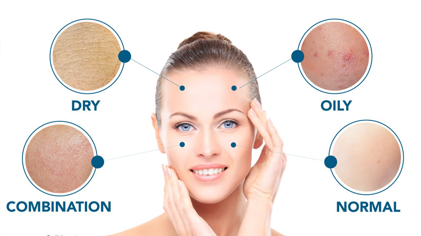
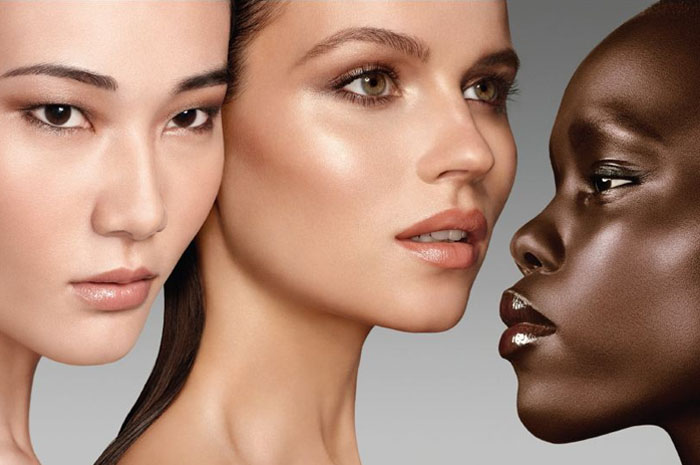
When determining skin type, two different conversations quickly surface. There is the more beauty-focused skin type discussion that refers to how our skin feels, how much oil it produces and how it reacts to products (think: normal, dry, oily, etc.) and then there is the skin type discussion that focuses on how our skin reacts to the sun and its susceptibility to skin cancer symptoms (think: fair, medium, dark, etc.) Both branches of the skin type conversation root back to our genetics.
We are born with the skin we have and cannot do much to change its natural properties. That’s why understanding the nature of our skin — in all its forms — is important for keeping it healthy and looking good.
Below, we take a look at the two kinds of skin type and explain how you can determine and care for your individual type. For our purposes, we are going to refer to the two branches as “sun exposure skin type” and “beauty skin type.” But even though we are dividing them, it’s important to remember that every feature of our skin is intimately connected, and the way our skin reacts to beauty products may be closely related to how it responds to the sun.
Sun Exposure Skin Type
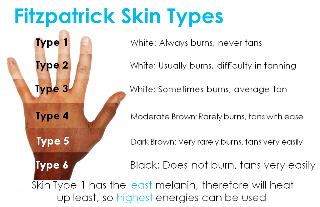
The most widely used system for determining skin type and how it will react to the sun is the Fitzpatrick scale. Created by Harvard dermatologist, Thomas B. Fitzpatrick, in 1975, the scale was developed to determine the response of skin types to ultraviolet light. Dermatologists realized that looking at hair and eye color alone as a way to predict skin sensitivity to the sun was ineffective. They made this scale inclusive by looking at how patients’ skin reacted to the sun and cataloguing responses into a general scale.
The scale is divided into six different skin types. Check out the different indicators for each type to determine which category your skin falls into.
Once you know where you fall on the scale, it’s important to know how to care for your skin type:
Type 1: Type 1 skin types need to be the most careful in the sun. With the least amount of melanin in your skin, you are the most vulnerable to skin cancer. Try to use a sunscreen with a SPF of 30+ and seek shade whenever you are out in the sun. Be sure to check your skin head-to-toe each month for suspicious spots or moles.
Type 2: Type 2 skin types are also very susceptible to skin cancer and should practice precaution in the sun. While your fair skin will tan occasionally, it is good to wear a SPF of 30+ when in the sun and avoid being in direct sunlight for extended periods. Be sure to conduct regular, head-to-toe skin checks each month as well.
Type 3: As a type 3, you may burn at the beginning of the summer but tan easily afterwards. Your medium to olive skin tone is more protected than types 1 & 2 but it still requires a strong SPF of at least 30+ to stay safe. Try to check your skin for moles and spots every month, or at least every three months, to prevent skin cancer.
Type 4: Your medium-brown skin tans easily and rarely burns. You are less likely to get skin cancer, but that doesn’t mean you shouldn’t protect your skin from the sun’s harmful UV rays. Use an SPF of at least 15+ every day and avoid direct sun exposure.
Type 5: Type 5 skin tans very easily and seldom burns. Skin cancer is more rare for your skin type, but when skin cancer does occur it is usually detected at a later and higher-risk stage, usually on areas not directly exposed to the sun such as the palms of the hands. Use an SPF of 15+ while outside, and perform skin checks at least every three months to ensure that nothing goes undetected.
Type 6: As the darkest skin type, you almost never burn and tan very easily. But just because you never burn, doesn’t mean you are free of skin cancer risk. Still practice caution and use an SPF of 15+ to prevent damage to skin cells. Like Type 5, you are also more at risk for skin cancer in less exposed skin areas. Check your skin often for any changes or suspicious moles or spots.
To get a more accurate idea of which type you are, take the Fitzpatrick skin type test.
Beauty Skin Type
Now that you know how your skin fairs in the sun, it’s time to identify the other factors that define your skin, specifically the skin on your face. While most skin exhibits features of multiple categories, having an idea of how your skin normally behaves will help you know how to care for it and which products to use.
To gain the best idea of which skin type you have, wash your face first and let it dry. Don’t put any products on it for an hour and see how it acts naturally. This will give you a good indication of its true nature.
Normal skin type
Normal skin is not too dry or oily; it falls in that desirable in-between place. Your skin normally has an even tone and a soft texture, with little flakiness. You may get an oily T-zone (the central area of your face including the chin, nose and the part of your forehead above your eyebrows) in hot weather, but generally, this area is oil-free. Lighter lotions and serums are ideal for your skin type because you don’t need much heavy product to keep your skin feeling great.
Dry skin type
Dry skin is characterized by small pores and an overall feeling of tightness. It often has more visible lines, less elasticity and a duller complexion. Moisture is key to caring for dry skin. Use lotions or creams to nourish your skin cells. If your skin feels dry but you still get breakouts, then you don’t have truly dry skin. Your skin may be feeling dry from the products you’re using.
Skin care routine for dry skin
Oily skin type
Oily skin tends to have larger pores, and a shiny, thicker feeling complexion. Blackheads and pimples are more common with this skin type. If you blot your face with a tissue and oil stays behind, then it’s likely that you have oily skin. Cleansing the face often and avoiding heavy creams and emollients is advised for minimizing the appearance of oil.
Oily Skin Type: how to treat and what are the symptoms
Combination skin type
While most of us have combination skin to some degree since there are more sebaceous glands around our nose, this skin type is marked by a consistently oily T-zone with dryness in other areas of the face. This is the most common skin type, and people with combination skin should consider using different products for different areas of the face to keep the skin balanced.
Additional Skin Type Factors
There are a few other features that contribute to skin type. It’s good to take these into account as well when figuring out what kind of skin you have.
Sensitive Skin
Sensitive skin reacts easily to products and can breakout in rashes or become itchy. This is more likely in fair to medium skinned people but any skin type can have sensitivities. If you have sensitive skin, avoid harsh products and search for products that don’t clog or irritate the skin.
Acne-Prone
Skin can also be acne-prone. Acne of all kinds can occur at any age and on any area of the skin. Oily skin types are more likely to have acne but it can occur with all skin types. Consult a dermatologist to find an acne product and care routine that works for your unique skin problems.
Understanding all of the features of your skin may seem like a lot of work, but it is the best thing you can do to care for it properly. Once you understand how your skin responds to different elements, you can zero in on what products, treatments and safety precautions are ideal for you.
What kind of skin do you have? What are the best ways you’ve found to care for it? Let us know in the comments.
Tanning beds and other artificial tanning machines are harmful for everyone, regardless of skin type. Some research suggests that people who use tanning machines before age 35 years are 75 times more likely to develop melanoma in their lifetime.
Your risk of sun damage is also higher if you live near the equator. The closer to the equator you are, the more intense the sun’s rays are, so being vigilant about sun protection is crucial.
Everyone should apply sunscreen daily to receive maximum protection. Here’s what else you should know about your skin and how to protect it based on your skin type.
Types 1 and 2
If your skin type is 1 or 2, you have a high risk of:
sun damage
skin aging from sun exposure
melanoma and other skin cancers
You should follow these tips to protect your skin:
Use a sunscreen with an SPF of 30 or greater. Limit your sun exposure and seek shade whenever you’re out in the sun. Wear a hat with a wide brim to protect your head and face. Wear UV-blocking sunglasses. Wear protective clothing with a UPF rating of 30 or higher if you plan to be in direct sunlight for extended periods. Check your skin from head to toe each month. Have an annual skin checkup with a doctor.
Types 3 to 6 If your skin is type 3 to 6, you still have some risk of skin cancer from sun exposure, especially if you’ve used an indoor tanning bed. You should still use sun protection even though your risk is lower than people’s with type 1 or 2 skin.
The Skin Cancer Foundation notes that African-Americans who have been diagnosed with melanoma usually are often diagnosed at a later stage, contributing to a poorer overall outlook.
For maximum protection, you should follow these tips:
Limit your sun exposure.
Wear a hat with a wide brim to protect your head and face.
Wear UV-blocking sunglasses.
Wear protective clothing if you plan to be in direct sunlight for extended periods.
Wear sunscreen with an SPF of 15 or greater.
Check your skin from head to toe each month. Pay careful attention to any strange growths. Acral lentiginous melanoma is the dominant form of melanoma among darker-skinned people. It appears on parts of the body not often exposed to the sun. It’s often undetected until after the cancer has spread, so make sure you check all areas of your body.
Have an annual skin checkup with a doctor.
When to get screened
If you’re at an increased risk of skin cancer, you should have regular skin exams. Talk to your doctor about how often you should come in for a screening. Depending on your individual needs, skin screening could be more frequent than your annual checkup.
People at increased risk of skin cancer include those who have:
personal or family history of skin cancer
Fitzpatrick skin type 1 or 2
a compromised immune system
You can also talk to your doctor about how and when you should do your own skin checks.
About the Fitzpatrick scale
If you’ve ever tried to match foundation or concealer to your skin, you know just how tricky skin typing can be. Enter Fitzpatrick skin typing, a scientific skin type classification. Though this form of skin typing won’t help you find your perfect shade, it can tell you just how much shade you should get on sunny days.
Developed in 1975, the system classifies skin type according to the amount of pigment your skin has and your skin’s reaction to sun exposure. This information can help predict your overall risk of sun damage and skin cancer.
Once you know your risk level, you can arm yourself with the tools you need to protect your skin. Read on to learn your Fitzpatrick skin type, what sun protection you should use, and more.
Some days our skin simply won’t cooperate and we have no time to deal with it. All we want when this happens is a quick solution that can get us on our way and doesn’t require an extra trip to the store. For those days, it’s time to turn to a home remedy. You know, those solutions that are passed down from generation to generation and can usually be made with a few ingredients from your pantry.
Below we list three of the most tried and true home remedies for getting clear skin in a pinch.
List for getting clear skin in a pinch
Apple Cider Vinegar
The benefits of apple cider vinegar (ACV) are being touted everywhere these days, and for good reason. This ingredient is anti-bacterial and antiseptic, which means it kills bacteria that can clog our pores and cause breakouts. It is made from apples that undergo a double fermentation process. This process makes ACV rich in many vitamins and minerals as well as an acid called malic acid. Malic acid has antibacterial, antifungal and antiviral properties, adding to ACV’s ability to fight acne and ward off pimples. ACV also helps balance the pH (acidity level) of our skin. Since our skin is naturally acidic, many basic soaps and cleansers can disrupt its balance, which, can in turn, inadvertently cause more oil production and breakouts to occur. The slightly acidic nature of ACV helps restore balance to our skin.
How to use:
It’s always advised to dilute ACV before applying to your skin because it can be quite strong. Mix one-part ACV and one-part water to make an easy and effective skin toner. Use like your normal toner, avoiding the areas around the eyes, or leave on and rinse off after a few minutes if your skin is more sensitive. As ACV can be too intense for many people at first, be sure to do a spot test before using. Dilute the ACV with more water if you experience a mild reaction. You can always up the strength over time as your skin adjusts.
Honey
Honey has been used for centuries as a way to heal and soothe the skin. Its antimicrobial and antibacterial properties make it ideal for cleansing the skin and getting rid of pimples. Honey also makes a great cleanser because it naturally helps our skin retain moisture, keeping it soft and reducing wrinkles.
How to use:
Buy any type of raw honey (although many people say Manuka honey offers superior antibacterial and healing properties) and use it as you would your normal face cleanser. You can also use it as a spot treatment, dabbing it on to your pimples and letting it sit for 10-15 minutes before rinsing off.
Tea Tree Oil
Tea tree oil is made from the Australian paperbark tree (Latin name: melaleuca alternifolia). This tree has been used for centuries by Australian aborigines for a range of healing purposes and its benefits have recently caught on all over the world. It has natural antibacterial, antifungal and antiviral properties and it can be used to treat a wide variety of skin ailments. Because of its ability to kill bacteria, it is especially helpful for treating acne and breakouts.
How to apply:
Before using tea tree oil, dab a few drops on a cotton swab, wipe it on your inner arm and wait a few minutes to see if your skin reacts. If nothing happens, it is safe to use undiluted on your skin. If you experience a mild reaction, you’ll want to dilute it by 50 percent with water. Once you know your tolerance, use it like an acne treatment. Wash your face as usual and use a cotton pad to apply the oil directly to your breakout, no need to rinse it off. Do this in the morning and evening. You can also use it as a toner by adding four drops of tea tree oil to one cup of water and applying it once a day. Other recipes advise for every 25 ml of water to add 10 drops of tea tree oil. Find a strength that works for you. You can add other essential oils as well for fragrance and additional benefits.
The majority of skin lesions are benign, but when a new lesion or mark appears on our skin, it can be difficult to tell whether it is dangerous. If you have any suspicions about a mark, mole or lesion, you should ask your doctor to check it. Nevertheless, it is useful to know how the common skin lesions look like to be able to recognise them.
In this post, we explain all about the most common skin lesions (with photos) and their main characteristics.
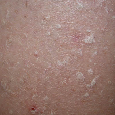
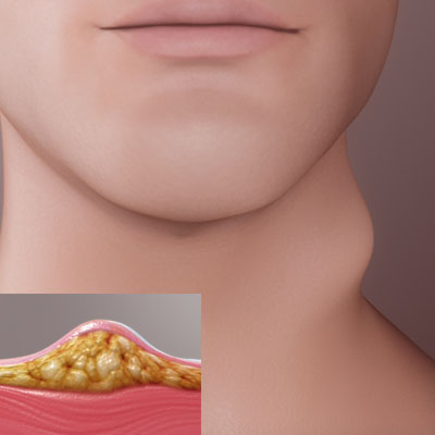
Seborrheic Keratosis
Seborrheic Keratosis, sometimes called senile wart, is a non-cancerous condition that occurs as a light brown, black or tan growth on the surface of the skin. These are usually harmless but may sometimes get irritated or be aesthetically unappealing. They can be removed, if necessary.
Common characteristics:
· These lesions are slightly elevated
· They are waxy and scaly
· They can appear in all areas of the body except for the palms and soles
· They often occur later in life and usually in multiples
· Many times, it has a stuck, “pasted on” appearance
Dermatosis papulosa nigra
Dermatosis paulosa nigra is a condition that occurs mainly in darker skin types and usually starts to form in adolescence. The lesions are small, darkly pigmented papules that are harmless and generally don’t require treatment.
Common characteristics:
· It appears mainly on cheeks and foreheads
· It often occurs in multiples
· This lesion has a smooth surface
· It appears most likely in an old age
Stucco keratosis
Stucco keratosis is another harmless skin condition among common skin lesions with the following characteristics:
· It consists of small, white-gray papules
· It appears mostly on the ankles or feet
· Men are more likely to have it
· It is more common among fair-skinned individuals
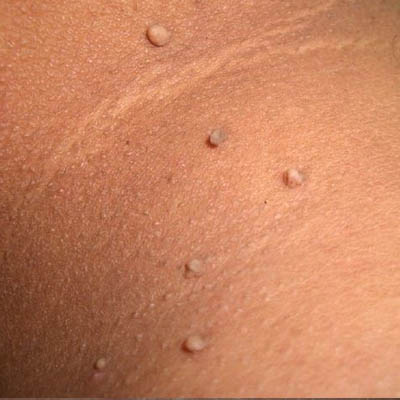
Skin tags (acrochordons)
Skin tags or acrochordons are soft skin growths where a narrow papule sticks out of the skin from a short piece of flesh like a tag.
Common characteristics:
· They are fleshy
· They often occur on the eyelids, neck, groin or armpit
· They can become irritated if twisted or rubbed a lot
· They are harmless but can be removed for cosmetic reasons
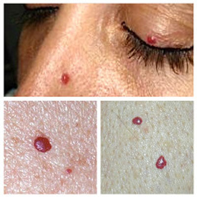
Cherry angiomas
Cherry angiomas are red papules filled with blood vessels made up of capillaries at the surface of the skin.
Common characteristics:
· They usually develop after the age of 40 and increase in number over time.
· They occur in higher concentrations on the trunk of the body.
· They may resemble melanoma when they bleed or clot.
· They do not require treatment, unless for cosmetic reasons.
Dermatofibroma
Dermatofibroma is a benign skin tumor that appears as a firm, round, brownish to red-purple growth usually found on the legs.
Common characteristics:
· It feels like a hard lump under the skin
· When squeezed, it dimples since the lesion is tethered to lower layers of the epidermis.
· It initially has a red color, later changing to brown
· It is dome-shaped
· It usually has a darker peripheral rim
· It is often a result of a prior injury
Solar lentigo
Solar lentigos, also known as “sun spots” or “age spots,” are marks on the skin from sun damage that are not cancerous. Although no treatment is required, patients are often at an increased risk for skin cancer and need to exercise precaution.
Sebaceous hyperplasia
Sebaceous hyperplasia is a skin condition that occurs when sebaceous gland on the skin is enlarged.
Common characteristics:
· It appears as small, skin-colored to yellow papules with a central indent
· These papules often occur on the forehead and central face
· They can often resemble basal cell carcinoma, but they rarely bleed or crust
· Removal is not necessary, unless for cosmetic reasons
Epidermal inclusion cyst (EIC)
An epidermal inclusion cyst is a common skin cyst, sometimes called sebaceous cyst, even though it arises from hair follicles, not oil glands. The cyst contains a foul-smelling, cheese-like substance formed from degenerating keratinocytes. They can become red and aggravated if the substance enters the dermis, an occurrence which is often mistaken for an infection. Asymptomatic cysts don’t require treatment. Excision can be used to remove a cyst if desired.
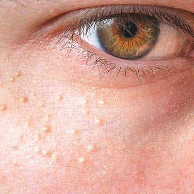
Milia or tiny epidermoid cysts
Milia or tiny epidermoid cysts is a condition where small 1-2 mm white to yellow papules occur underneath the surface of the skin.
Common characteristics:
· These cysts can occur in all ages
· They can be extracted without scarring
· They are fixed and long-lasting
· They often occur on the cheeks, eyelids, forehead, and genitalia.
Lipoma
Lipomas are collections of fat under the skin. These common skin lesions are soft and mobile benign tumors that usually stop growing when they reach a few centimeters in diameter. Treatment is surgical and considered elective.
Common characteristics:
· They often appear on the trunk, arms, or thighs
· They tend to occur in multiples in early adulthood
A lipoma is a slow-growing, benign, adipose tumor, often found in the subcutaneous tissues. Lipomas may also occur in deeper tissues such as the intermuscular septa, the abdominal organs, the oral cavity, the internal auditory canal, the cerebellopontine angle and the thorax or any part of the body where there are fat cells. However, they are mostly asymptomatic, unless they press on a nerve causing pain, or rarely develop in the gut wall leading to blockage.
Several factors may increase your risk of developing a lipoma, including:
Middle ageLipomas are most common in the age group of 40 to 60, and typically rare in children.
Other disordersPeople with Dercum’s disease (painful lumps on the trunk, shoulders, arms and legs), Cowden syndrome (hamartomas present in the skin, mucous membranes, thyroid gland, and breast tissue) and Gardner’s syndrome (intestinal polyposis, cysts and osteomas), have an intensified risk of developing multiple lipomas.
GeneticsThere is an uncommon autosomal dominant condition called familial multiple lipoma in which groups of fat cells occur under the skin and then produce multiple symmetrical lumps. This runs in families.
Treatment for a lipoma usually isn’t necessarily unless it starts to bother. It is usually carried out in the following ways:
SurgeryMost lipomas are extracted surgically. The side effects are scars and bruises which may possibly remain. Recurrences are not common.
LiposuctionSince lipomas are fat-based, this procedure can work well to reduce its size. A needle and large syringe is used to remove the fatty tissue.
Steroid injectionsSteroid injections may also be used right on the affected area. Their use before the surgery is being studied. This treatment shrinks the size of lipoma but usually doesn’t remove it. The reduced lump can then be removed by surgery.
A cancerous, fatty tumor is termed as liposarcoma. Liposarcomas grow rapidly and are usually painful. There is a very little chance that a lipoma turns out to be a malignant liposarcoma and the suspected lump is examined through a biopsy, MRI or CT scan.
Images courtesy of the American Academy of Dermatology
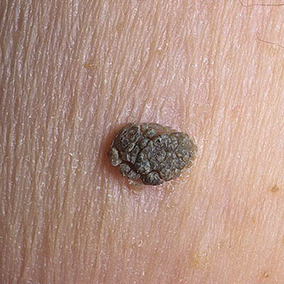
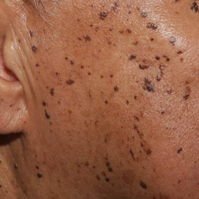
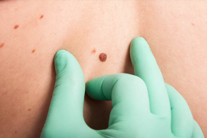
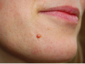
While for many of us moles are just brown spots on our body we may not pay much attention to, they come in various shapes, sizes, and forms that can tell us important things about our skin health. Understanding all types of skin moles helps us identify any suspicious spots for skin cancer and keep our skin healthy.
Want to keep your skin healthy? Use Skinive to check your moles for signs of skin cancer and get an instant risk indication.
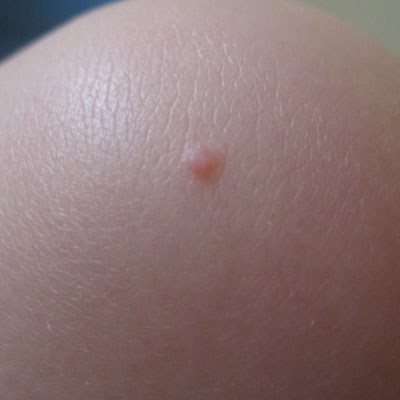
First of all, what is a mole?
A mole or nevus is a dark spot on our skin comprised of skin cells that have grown in a group rather than individually. These cells are called melanocytes and are responsible for producing melanin, the pigment (color) in our skin.
Moles appear on our skin from sun exposure (ultraviolet radiation), or we are born with them. Although the number of moles varies from person to person, fair-skinned people generally have more moles due to lower amounts of melanin in their skin. The average number of moles for adults is between 10 and 40. Moles can even come and go with hormonal changes such as pregnancy or puberty.
Most people develop more moles on their skin naturally with age and sun exposure, and most of the time these are harmless. However, we need to conduct skin checks regularly (recommended monthly, especially there is a family history of skin cancer, or at least every three months) to check whether they have changed.
Download Skinive now to check your skin and get instant risk indication of skin cancer.
Types of skin moles
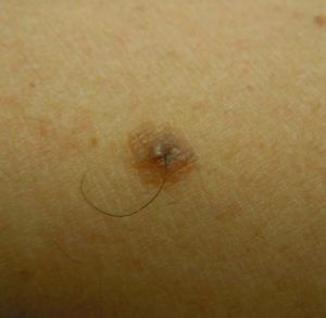
Not all moles are created equal. Here’s a quick guide to mole types and what they mean for your skin. We categorised moles based on time of development, placement on the skin, and typical or atypical symptoms. Based on this categorisation, moles can be described by multiple classifications. For example, you may have a common acquired junctional nevus or an atypical congenital nevus.
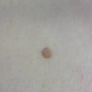
Common
A common mole is usually about 5-6 mm large, has distinct edges, a smooth, dome-like surface, and pigmentation. Common moles are found on skin regularly exposed to the sun and can potentially but rarely turn into skin cancer.
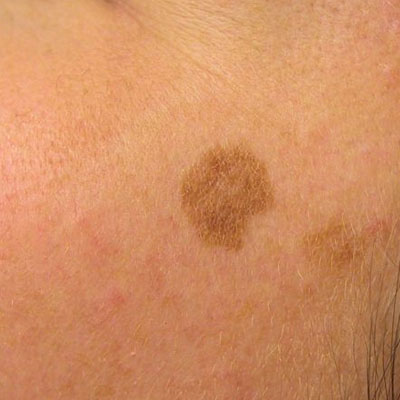
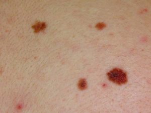
Atypical
Atypical (dysplastic) melanocytic naevus
Atypical moles, or dysplastic nevi, are moles that exhibit irregular symptoms. They usually have fuzzy or blurry borders, vary in colour, are larger than most moles, and have both flat and raised components. While dysplastic nevi share a lot of the same signs of pre-cancerous or cancerous moles, most dysplastic nevi are benign. However, a person with many dysplastic nevi has an increased risk for skin cancer. Regular self-examinations are important to detect changes in these types of moles.
What is Skin Cancer?
Mole types by time
Congenital
Congenital moles, also known as congenital nevi, are moles that appear at birth or within the first year of a baby’s life. They are caused by melanocyte cells in the dermis (middle layer of skin), epidermis (outer layer of skin), or both. These types of moles can range in size and are sometimes referred to as birthmarks. Congenital nevi pose some risk of developing into melanoma later in life and should be monitored as you enter adolescence and adulthood.
Acquired
Acquired moles are moles that appear during childhood and adulthood. Most of these moles are benign and pose no risk, although sometimes they can turn into cancerous moles with age. This type of mole is the most common, and repeated sun exposure usually causes it.
Mole types by location
Junctional Melanocytic Nevi
Junctional melanocytic nevi are moles that occur from an accumulation of melanocytes where the dermis and epidermis meet. These moles are typically slightly raised with regular borders and dark pigmentation, although they can range in color from tan to dark brown. People usually acquire these moles between childhood and early adulthood, because, as we age, it is common for melanocytes to migrate down to deeper layers of the skin.
Intradermal Nevi
Intradermal Nevi
Intradermal nevi are skin-coloured moles that often blend in with your surrounding skin. Their pigmentation is not as dark as junctional melanocytic nevi because they are located in the dermis or the middle layer of your skin. These moles usually develop in late childhood or throughout adulthood and are very common and usually benign.
Compound Nevi
Compound Nevi
Compound nevi show signs of both intradermal and junctional nevi, with melanocyte cells located in the dermis and dermo-epidermis junction. These moles usually have a central raised area with flat areas around the edges. They usually have distinct borders and even pigmentation.
Other mole types to note
Halo Nevi
Benign Formation – Halo Nevus
Halo nevi are raised moles surrounded by a ring of skin that has lost pigmentation due to inflammatory infiltrating cells. Doctors are still trying to understand why this reaction occurs, but if appropriately diagnosed, these moles are benign and require no treatment unless for cosmetic reasons.
How to know if your mole is safe
Moles that are considered “safe”, or not at risk for cancer, generally have a few common features.
They usually resemble common moles and:
have neat edges,
are smooth or dome-shaped,
are around ¼ inch (6 mm) in diameter,
preserve the same shape, size or colour over time.
It is important to make an ‘inventory’ of your moles to recognize any changes that may occur over time. Understanding what is normal for your body is key to the identification of early skin cancer symptoms and in-time treatment.
Warning signs that it may be cancerous
Look for these indicators that your mole may be cancerous:
Changes in size (getting larger)
A change in shape (especially with irregular edges)
Colour changes (especially getting darker or exhibiting multiple shades)
A loss of symmetry (common moles will be perfectly round or oval and are usually symmetrical)
Itchiness, pain or bleeding (maybe even forming a scab)
Crustiness
Inflammation
Exhibiting three different shades of brown or black
A change in elevation (thickening or raising of a flat mole)
If you notice any of these symptoms, contact a doctor to examine your mole.
Skin cancer pictures
ABCDEs of Moles
Remembering all of the things to look out for during a skin check can be difficult: this is why doctors advise using the ABCDE method to make things simpler. Check your skin for these signs during self-examinations:
Asymmetrical – the mole is distinctly asymmetrical Border – the mole has uneven borders Colors – the mole contains at least two distinct colours Diameter – the mole is bigger than ¼ inch or 6 mm across Enlargement – the mole grows in size over time
In most parts of Europe and USA, people are currently enjoying plenty of sunshine as the summer is coming closer. But the sun hasn’t shown its full potential yet. So, how can you enjoy the heartfully warm days while keeping your skin healthy? Here are five tips on how to keep a healthy skin in the summer, without missing out on the pleasures of sunlight.
1. Be cautious with tanning
Tanned skin is often considered a sign of health and attractiveness, but in reality, it is a sign of skin damage. Research has shown that the continuous damage to skin cells from exposure to UV light increases the likelihood of skin cancer and melanoma. In fact, 90% of melanomas are estimated to be caused by UV exposure.
2. Wear sunscreen
We can’t stress this enough: the sun is lovely, but it can be harsh on your skin. Wear sunscreen every time you come across direct sunlight. For maximum protection from UV light, apply sunscreen with SPF 30 or more, at least half an hour before you get exposed to the sun. You will still tan while protecting your skin.
3. Drink water
During hot days, your body sweats more often to cool down its temperature, which results in constant loss of fluid. Your skin cells moisturize your skin using their natural chemicals, among which water is the key ingredient. Any loss of the body’s water volume in a day can lead to skin dehydration. To avoid this, drink water throughout the day. Make sure you always have a bottle of water with you, even when you are not exposed to direct sunlight, to keep a hydrated and healthy skin in the summer.
4. Seek some shade
When temperatures are rising, it is important not to stand under the sun for too long. The UV rays are most harmful between 11 am and 4 pm, so make sure you seek some shade during this time to protect your skin from possible damages.
5. Check your skin regularly
Most skin diseases are entirely treatable when diagnosed at an early stage. In addition to your daily beauty routine, it is always recommended to perform regular checks on your skin. You can do this easily and comfortably with SkinVision directly from your smartphone. Try it now!